Anatomy Of Anterior Chest Wall / Thoracic Wall Atlas Of Anatomy - Swensen for orientation, we show here the anterior chest wall and we show the pectoralis major which forms the anterior axillary fold, we point out now.
Anatomy Of Anterior Chest Wall / Thoracic Wall Atlas Of Anatomy - Swensen for orientation, we show here the anterior chest wall and we show the pectoralis major which forms the anterior axillary fold, we point out now.. Anatomy, breast, axilla, chest wall, metastatic sites. The azygos vein courses anterior to the spine, either behind or to the right of the esophagus, until it arches anteriorly to join the posterior wall of the svc. We compared the sensitivity and specificity of bone scans and mri in assessing. Paired mammary glands, or breasts, are a distinguishing feature of mammals. Right and left scapular lines:
In this article we will focus on: Sometimes the lower, traveling on the upper. The capsule gains much support comes from the radiate ligament or anterior costovertebral ligament which attaches to the rib head anteriorly and to the sides of the articulating vertebral bodies and the. The interpretation of a chest film requires the understanding of basic principles. Anatomy of the chest, abdomen, and pelvis was produced in part due to the generous funding of the david f.
3 The Thorax Pocket Dentistry from pocketdentistry.com The chest wall has 10 layers, namely (from superficial to deep) skin (epidermis and dermis), superficial fascia. Anatomical landmarks that play an important role in clinical. Chest wall movement o anterior posterior diameter: Liszewski ricardo restrepo edward y. This first part covers the muscles of the anterior abdominal wall. Pump handle sagittal plane § attachment. I always check the chest when a patient complains of chronic neck pain or symptoms in the a patient s guide to rib joint pain anatomy where are the rib joints? Clinical anatomy students learn to use imaginary lines and bony landmarks on the front and back of the thorax to describe locations of the anatomical structures.
The interpretation of a chest film requires the understanding of basic principles.
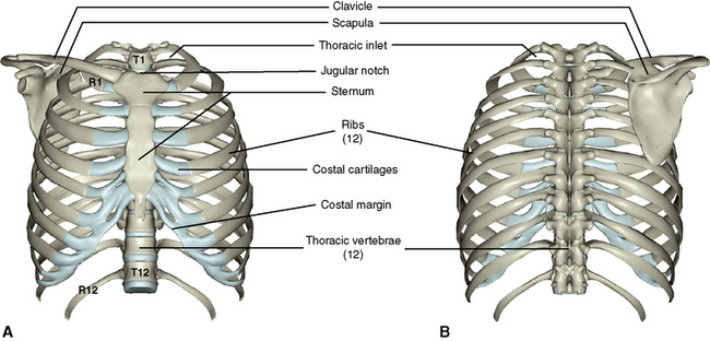
Ribs 3 through 9 are typical ribs as described earlier while ribs 1, 2, 10, 11, and 12 are atypical. The precordium is the region of the anterior chest wall immediately anterior to the heart. Sometimes the lower, traveling on the upper. It passes anteriorly and laterally. Simple, easy notes for quick revision of important questions. The chest wall is supplied by the posterior intercostal arteries arising from the aorta, the internal thoracic and the highest intercostals given off the the anterior intercostal arteries are sometimes two (inferior and superior) in the respective intercostal space; The capsule gains much support comes from the radiate ligament or anterior costovertebral ligament which attaches to the rib head anteriorly and to the sides of the articulating vertebral bodies and the. Anterior chest wall involvement is not infrequently observed within inflammatory arthropaties, particularly if one considers seronegative spondyloarthritides and sapho syndrome. 2 name the planes used for dividing abdominal cavity into regions. Bone scan is sensitive to acw involvement, while magnetic resonance imaging (mri) detects early alterations in spa. Anterior chest wall (acw) involvement is difficult to evaluate in patients with spondyloarthritis (spa). I always check the chest when a patient complains of chronic neck pain or symptoms in the a patient s guide to rib joint pain anatomy where are the rib joints? .12 photos of the anatomy of skull anatomy of cow skull, anatomy of newborn skull, anatomy of skull animation, comparative anatomy of vertebrate skull female pelvis, diagram of anterior female pelvis image, diagram of female pelvis and reproductive tract, hip diagram female, human anatomy.
Cc sternum ribs attached to costal. 1 enumerate the layers of anterior abdominal wall. In general, the anterior abdominal wall has nine layers (from superficial to deep) Anatomy of the chest and the lungs: 3 what are the vertebral levels of important.
Options For Flap Plasties Of The Anterior Chest Wall Download Scientific Diagram from www.researchgate.net We compared the sensitivity and specificity of bone scans and mri in assessing. The bony skeletal part of the thoracic wall is the rib cage, and the rest is made up of muscle, skin, and fasciae. The muscle covers the anterior chest wall medially and flattens to a 5 cm long tendon which inserts into the proximal anterior humerus. Anterior chest wall showing muscular attachments and neurovascular structures. Pectus excavatum is a congenital deformity of the ribs and the sternum producing a concave appearance of the anterior chest wall. The capsule gains much support comes from the radiate ligament or anterior costovertebral ligament which attaches to the rib head anteriorly and to the sides of the articulating vertebral bodies and the. Simple, easy notes for quick revision of important questions. Understand the importance of active recall in learning anatomy and start using this technique to ease your work, improve retention and save time!
Clinical anatomy students learn to use imaginary lines and bony landmarks on the front and back of the thorax to describe locations of the anatomical structures.
3 what are the vertebral levels of important. In general, the anterior abdominal wall has nine layers (from superficial to deep) The anterior wall is formed by the aponeuroses of the external oblique, and of half of the internal oblique. In this article we will focus on: Sometimes the lower, traveling on the upper. The bony skeletal part of the thoracic wall is the rib cage, and the rest is made up of muscle, skin, and fasciae. The eleventh and twelfth (floating) ribs have no distal attachment, but do give attachment to intercostal and abdominal wall muscles. Lee introduction the chest wall encases and protects the vital structures within the thoracic cavity. Anterior chest wall (acw) involvement is difficult to evaluate in patients with spondyloarthritis (spa). Anatomy of lung segmental anatomy of lung lateral view on a normal lateral view the contours of the heart are visible and the ivc is seen entering the right atrium. Ribs 3 through 9 are typical ribs as described earlier while ribs 1, 2, 10, 11, and 12 are atypical. Cc sternum ribs attached to costal. The serratus anterior, as its name suggests, consists of multiple muscle slips that run along the anterolateral chest wall (see figure 1 for surface anatomy).
Week chest wall (thoracic cage) anatomy component overview sternum manubrium body xiphoid process ribs to costal true ribs: Simple, easy notes for quick revision of important questions. The pectoralis minor is much smaller, and arises from the anterior ends of the 3rd to 5th ribs, beneath pectoralis major. Anatomyzone is youtube's most highly subscribed anatomy channel, with video tutorials on all areas of anatomy. The capsule gains much support comes from the radiate ligament or anterior costovertebral ligament which attaches to the rib head anteriorly and to the sides of the articulating vertebral bodies and the.
Chapter 14 General Principles Of Chest Trauma Operations Anesthesia Key from i0.wp.com The capsule gains much support comes from the radiate ligament or anterior costovertebral ligament which attaches to the rib head anteriorly and to the sides of the articulating vertebral bodies and the. Chest restrictions can contribute to numerous problems throughout the body. The muscle covers the anterior chest wall medially and flattens to a 5 cm long tendon which inserts into the proximal anterior humerus. Chest wall movement o anterior posterior diameter: The chest wall is supplied by the posterior intercostal arteries arising from the aorta, the internal thoracic and the highest intercostals given off the the anterior intercostal arteries are sometimes two (inferior and superior) in the respective intercostal space; Cc sternum ribs attached to costal. Right and left scapular lines: The chest wall is the structure that surrounds the vital organs within the thoracic cavity and consists of skin, fat, muscles, and bone (rib cage).
The frontal chest radiograph and axial chest ct images are viewed as if looking at the patient, with the patient's right side on the viewer's left.
It passes anteriorly and laterally. The anterior wall is formed by the aponeuroses of the external oblique, and of half of the internal oblique. The bony skeletal part of the thoracic wall is the rib cage, and the rest is made up of muscle, skin, and fasciae. In general, the anterior abdominal wall has nine layers (from superficial to deep) Anterior chest wall (acw) involvement is difficult to evaluate in patients with spondyloarthritis (spa). Anatomy of the chest and the lungs: The azygos vein courses anterior to the spine, either behind or to the right of the esophagus, until it arches anteriorly to join the posterior wall of the svc. A series of 31 fresh cadaver injection, dissection and radiographic studies were undertaken to define the vascular architecture of the anterior chest wall and to correlate the findings of previous writers in this area. The pectoralis minor is much smaller, and arises from the anterior ends of the 3rd to 5th ribs, beneath pectoralis major. Run parallel with the midvertebral line but pass through the inferior angles of the scapulae. The first rib is a short, flat rib that is much wider and more curved than those previously described. Anatomy, breast, axilla, chest wall, metastatic sites. Feeling overwhelmed by the anatomical details of heart valves?
Muscles of the anterior chest wall pectoralis major o the sternal part of the sternocostal head of the pectoralis major originates from the manubrium breast anatomy the breast extends from the 2nd to the 6th rib and the axillary tail projects into the axilla transversely, it extends from the lateral border of anatomy of chest wall. Ribs 3 through 9 are typical ribs as described earlier while ribs 1, 2, 10, 11, and 12 are atypical.


0 Comments